Plant Cell Diagram: Structure, Biological Function and Key Processes
Understand plant cell diagram with clear definitions, examples, scientific context, and notes on common misunderstandings.
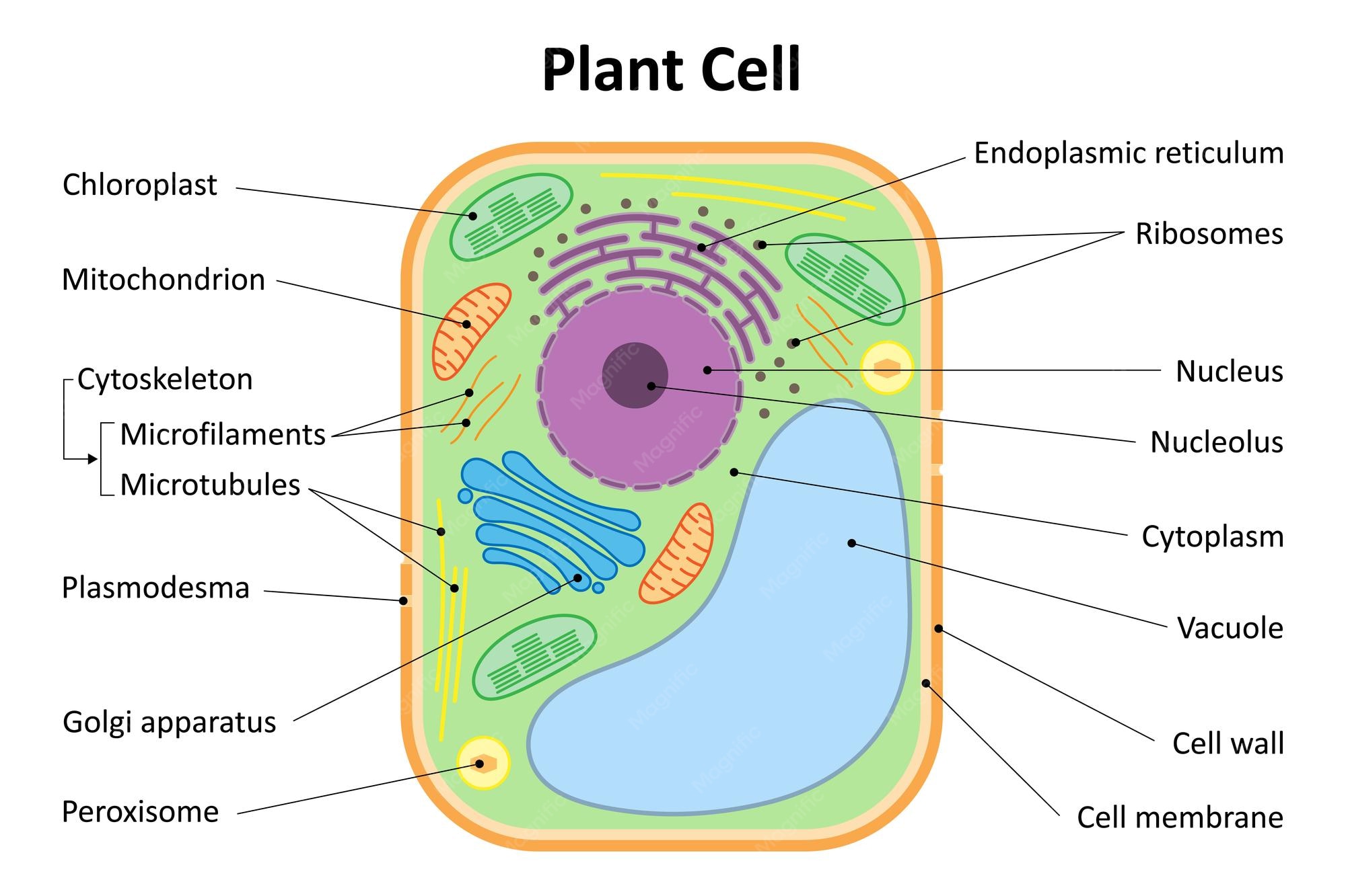
Plant Cell Diagram: Organelles, Labels and Functions
A plant cell diagram typically identifies the cell wall, plasma membrane, cytoplasm, nucleus, vacuole, and often chloroplasts.
A useful way to understand this topic is to separate the underlying idea from a worked example and from the limits of the explanation. Biological explanations connect structures with processes and inherited information. What a diagram shows at one scale may not describe every cell type or life stage.
How it works
The cell wall provides support, the membrane controls exchange, and chloroplasts capture light energy for photosynthesis in cells that contain them.
The mechanism matters because it explains the observation rather than just naming it. The pattern becomes clearer when the structures, context, and evidence are considered together.
An example in context
A typical leaf cell diagram may show a large central vacuole and green chloroplasts, while many plant root cells lack chloroplasts.
This example is best treated as a specific case, not an all-purpose rule. Consider which features are broadly shared and which depend on species, historical setting, or individual circumstances.
What the term should not be confused with
Plant and animal cells share many eukaryotic structures, but plant cells commonly have a cellulose-rich wall and plastids.
These boundaries help keep this page focused. Related subjects may overlap, but a shared word or superficially similar appearance is not sufficient evidence that two concepts are equivalent.
Why the distinction matters
Microscopy, molecular experiments, sequencing, and comparative observations provide complementary ways to examine cells and heredity. No single simplified drawing establishes every mechanism.
For readers, the practical lesson is to ask what is directly observable, what is inferred from a model, and which further evidence would help separate competing explanations. In this topic, the example above illustrates why that difference can matter.
Common mistakes and limitations
Educational diagrams are simplified and are not universal blueprints. Cell types, proportions, and organelles vary across plant tissues.
In biology, patterns often depend on organism, tissue, and environment. A classroom example should not be assumed universal without additional evidence.
Frequently asked questions
What is Plant cell diagram? A plant cell diagram typically identifies the cell wall, plasma membrane, cytoplasm, nucleus, vacuole, and often chloroplasts.
What is a clear example? A typical leaf cell diagram may show a large central vacuole and green chloroplasts, while many plant root cells lack chloroplasts.
What is a common misconception? Plant and animal cells share many eukaryotic structures, but plant cells commonly have a cellulose-rich wall and plastids.
Continue exploring
Cell Biology and Genetics Explained — a broader topic overview.
Smooth Endoplasmic Reticulum — a focused explanation of a related concept.
Codominance — a focused explanation of a related concept.
